Technology
Eight years and over $5 million in research costs have
provided the answers needed for providing a quality health
management system that can be economically provided to the world's
economically disadvantaged people. The collaborative efforts of
scientists at the North Carolina Institute of Technology, University
of Kentucky and other research facilities have made this vision of
relieving suffering in the world and promoting world peace become
reality.
Below is the
genetic study which helped to develop specific technology for
stopping the cancer process. Other studies involving heart
disease, arthritis, and diabetes have revealed that these illnesses
can also be corrected and reversed. However, the unique
benefit of the technology is prevention and the global projects will
be emphasizing this aspect of the technology.
|
|
P53 AND
ITS ASSOCIATES P21, CDK2,
P27 & METASTASIS OF CANCER
|
| |
Dr. Robert Jones has
presented and has had accepted by the FDA the study
outlined below. He is the inventor of the Cavitat
ultrasound device used to help locate jawbone
cavitations that are responsible for the onset of
cancer. "Cavitations" are explained
below.
"If you want to find
out how
genes
affect other genes, you
have to find
out how proteins affect other
proteins."
Roger Brent
"If you
want to find out how proteins affect
other proteins,
you have to study inhibition
rates
by chemical
toxins."
Robert Jones
Researcher Robert Jones' Presentation. .
.
As
I have explored the causes of cancer it has become
apparent that the real cause of cancer is genetic
protein based, in other words, toxic inhibition of
proteins within the cell structure allows or encourages
a cell, or group of cells, to become
malignant. The International genome project
flatly states that all cancers are genetic and are
caused by one or more of 3
things:
1.
Radiation
2.
Chemical Toxins
3.
Spontaneity
I have
concentrated my efforts to uncover the cause of cancer
as chemical toxins as we have no way to control or
measure the rate of radiation exposure. I started
on the conquest of exploring chemical toxicity and its
relationship to protein inhibition several years
ago. In the process of my research, a huge amount
of toxins have been looked at. As we narrowed the
search to a protocol of daily or chronic exposure, at
the top of the list were compounds of known carcinogen
and neurotoxins, these toxins being classified as thio
ethers. The most significant of these thio ethers
is dimethyl-sulfide. Although small in amounts of
exposure, the average inhibition of ability to bind to
the cellular membrane came to a startling conclusion of
more than 90% inhibition as an average for all three
proteins.
To distinguish which toxins in particular, we
tested 36 lanes on Affinity Labeling
gels. Specifically, as we set the protocols
for this research project we used toxicity samples from
over 900 extracted root canal teeth as a composite and
over 4000 bone fragments obtained from biopsy samples as
a separate composite. Root canal toxins and
cavitation toxins were tested separately to determine
how each toxin individually inhibited the binding
ability of the protein. Establishing published
cellular weights (amounts) of these proteins, we
proceeded to inject Affinity Labeling gels with amounts
of human protein as to the stated amount found in each
individual cell. So, therefore, using toxins
extracted from human samples and human proteins, we were
able to exhibit extreme or severe inhibition of
these individual proteins by chronic exposure to these
toxins. We then ran additional lanes on the same
Affinity Labeling gels to determine the effects of blio
toxins (fungi) and also mercury from dental
amalgam. As you will note during my lecture, the
cavitation toxins from a composite of 100 or more
cavitations was much more toxic than root canal
toxins.
I'd like to show you in our basic DNA, we
have the chromosome ladder as illustrated in blue and
protein in green with amino acids in
yellow.
Notes in red are comments by NCIT that perhaps can clarify points for non-scientific readers:
 |
The inhibitions I will demonstrate will have serious effect on the protein's ability to function in the nucleus of the cell and the downstream effects of these inhibitions and how they affect the cellular functions will be noted.
Inhibitions does not mean damage.
You will see that when the condition is corrected, the proteins can restore their ability to protect you from tumors. This makes cancer curable and not just a remission state.
|
|
 |
I'd like to talk to you about each protein individually and about how each functions which has been established by the Science of National Genome Research Project.
|
|
The 4 proteins that we are going to look at are P54, P21, CDK2 and then we are going to look at a new publication from the University of Michigan published in the Journal of the National Cancer Institute.
We
will look at
the inability to bind P27 to P21 and demonstrate how it allows the cancers to metastasis.
Notice here that other studies from reputable organizations are involved in this research. |
| |
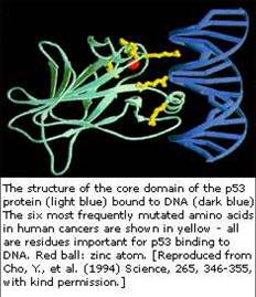
What is P53?
P53 is specifically a tumor suppressant protein.
It has been defined as a normal function of P53 to be antioncogenic. While type P53 proteins introduced into cells were foound to be growth
suppressand, P53 is found rarely in a tumor cell while it is very prolific in normal healthy cells.
When it is found in a malignant tumor, it is found sparingly and in an inhibited state as we will demonstrate and therefore its ability to bind in the cellular membrane is greatly reduced.
When P53 is normal or not inhibited, tumor growth or start is depressed.
The point being made here is that the P53 is a protein known to suppress tumor development.
In other words, it is an anti-cancer protein when it is allowed to function in a normal manner.
|
| |
Amino acids are an important class of organic compounds that contain both the amino (_NH2) and carboxyl
(_cooh) groups. Of these acids, 20 serve as the building blocks of proteins.
These amino acids are inhibited from binding to the chromosome ladder and or just one of the examples of damage incurred by these dental toxins.
The very building blocks of proteins are inhibited by the dental toxins.
This is one factor involved in the "wasting" process often seen in terminal cancer cases.
|
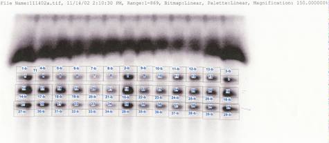
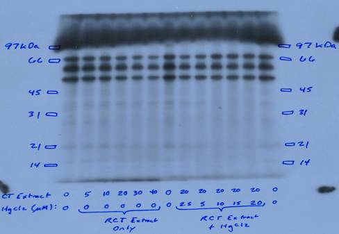
| This is the nucleotide protein gel before developing. |
 |
|
 |
If you will note, we have a graph that is printed out with a Hewlett Packard Phosphorescence measuring device as it measures radioactive light.
This is non-subjective analyzation of inhibition of proteins ability to bind and function.
|
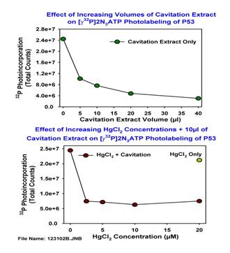 |
To the left vertically is the scale of light reading. The bottom from left to right is the amount of toxin injected into each protein extract.
They are shown in micro liters (ul).
|
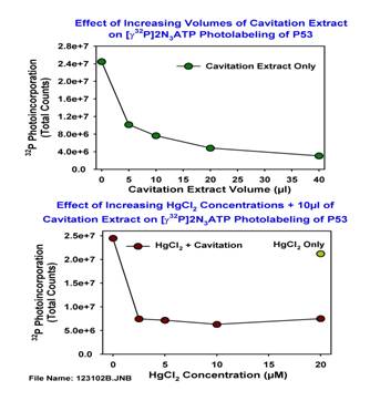 |
If you will note on P53 at 5 ul injection of the cavitation extract, the inhibition is already at 58.5%, any inhibition at over 12% will render functions to be ineffective.
What's interesting about this is that at 40 ul they are inhibited at about 85%.
A cavitation is a hole in the jawbone that is often (but not always) caused by poor dentistry.
Generally these osteonecrotic areas of bones are at the sites of extracted wisdom teeth, but can be at other extraction sites as well.
The researcher extracted tissue from these sites to perform the tests.
|
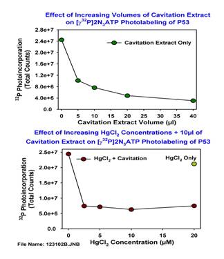 |
Then, we wanted to look at mercury amalgam vapors and what would they do to the inhibition aspects and we were surprised to find that mercury has very little effect in the binding ability to our DNA at this stage of toxicity.
In fact, photo labeling increased very slight when 10 ul was added to the cavitation toxins.
This is not to say that mercury by itself would not damage these proteins, but did not provide a synergistic effect.
So the condition was not exacerbated by the addition of the mercury.
We know mercury is dangerous, but it appears that it is not a direct contributor to the cause of cancer.
Mercury, of course, is well known for it ability to depress immune functioning, therefore, it contributes to the body's inability to fight the disease once in progress.
|
 |
Now we are looking at a graph of mercury from amalgams added to the Affinity Labeling gel.
As you will note, photo labeling of the protein P53 increased with increasing amounts of mercury.
So mercury is not the cause of inhibition at this stage.
|
 |
This slide shows gliotoxin concentrations produced in micro molar amounts.
At 5 um this protein showed 40.7% inhibition. This is a very severe inhibition at very low levels.
Even at very low levels of neurotoxins coming from the jawbones and dead teeth (such as a root canal tooth), severe inhibition can occur.
|
 |
Of all three of these proteins, P53 is the top dashed line with the circles.
As you note on the chart, P53 from root canal extract inhibits at 29% at 5 ul, continuing on at 40ul at 87.5%.
The interesting thing about these toxins is, it is estimated that a molar root canal produces 45ul each 24 hours!
All root canals are infected and some do produce more toxins than others.
Yes, root canal teeth!
The dentist saves a tooth and sets you up for cancer. There are NO GOOD ROOT CANALS regardless of what the American Dental Association says about root canals.
There is no evidence of any double blind study ever made on the safety of rooth canal procedures.
|
| |
P53 is shown now to be almost totally inhibited.
You must remember that Druckrey (Heidelberg) found these dental toxins to be the worlds most perfect carcinogen.
Creating an appearance and function of a malignant cell until the carcinogenic dose was reached.
Yes, you read it correctly--the world's most perfect carcinogen (a substance that is a known cause of cancer) is the dental toxins.
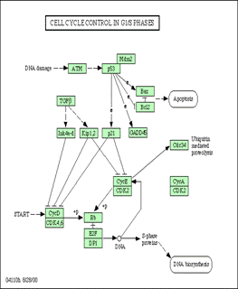
Now we are going to jump over P21 and go to CDK2.
|
 |
CDK2
|
 |
Keep in mind that CDK2 is a cyclin dependent kinase.
To function properly, CDK2 has to bind P21 after P21 has bound P53. We have already demonstrated that P53 is inhibited from binding to the cellular membrane of P21.
You are now seeing the development of the process and why cancer has been a very difficult disease to deal with.
Has the millions of dollars in research been given to scientists without the skills to investigate proteins?
|
| |
CDK2' main function in the GS1 phase is to control cellular mass size or cellular growth and if it is inhibited in ANY AMOUNT this function cannot occur.
|
 |
This slide, expressed by the Hewlett Packard light source, showing inhibition of CDK2 of cavitation extracts of 5ul
to be huge inhibition of 57.6% and that at 40ul, a whopping 89.5%.
The lower graph shows basically the same reaction to mercury as shown on P53.
|
 |
Now, we are looking at a graph of mercury from amalgams added to the Affinity Labeling gel.
As you will note photo labeling of the protein CDK2 increased with increasing amounts of mercury.
So, mercury is not the cause of inhibition at this stage.
|
 |
Now, back to P23.
|
 |
P21 or H-Ras is the smallest protein yet discovered, but it is the superman of proteins.
The primary function of P21 in the cellular structure is to control cell replication and apoptosis.
It is also interesting to note that the downstream function of P21 is to control the autonomic immune system.
In saying this, I will return later to this function.
The "superman" of proteins controls cell replication and apoptosis (cell death) which is an important factor in the cure.
Cancer cell death must occur to stop the cancer. Rather than destroy with chemicals, which destroys and damages even healthy cells, doesn't
it make more sense to optimize the P21 or H-Ras protein to do its job in the natural cell death of these cells? And, that is exactly what happens with the NCIT cure which does not involve chemotherapy.
|
 |
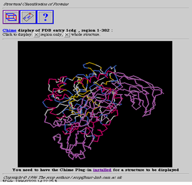
Protein P21 (Ras) & Protein P27 (Raf)
Stereo view of the association between ras-P21, a related protein rap1a, and downstream effector RAF.
This is a small part of the cellular signal transduction pathway. These images were obtained by docking ras-P21 and rap1a, each separately, to RAF, based on structural data obtained from x-ray crystallography, and minimizing until the RMS deviation was less than 0.1 angstroms.
|
| |
So, P21, an important regulatory protein in cell growth and differentiation is a GTPase called Ras.
There are 2 cellular switches on P21 that are activated by either GTP or as
in the second switch by GDP. When the cellular replication switch is turned on,
which
is regulated by hydrolyzing GTP, and the switch is turned off by the hydrolyzing GDP as in the second switch.
When GTP is bound to the first switch and is inhibited, it cannot hydrolyze GTP.
When P21 is unable to hydrolyze GTP the switch remains constant on. So, you have uncontrolled cell replication.
The second switch can not glycolyze GDP due to inhibition allowing the cell replication.
The second switch can not glycolyze GDP due to inhibition allowing the cell replication cycle to remain full on.
The full purpose of the second switch is to turn cell replication off and it cannot function in this capacity due to inhibition.
The second switch must be activated by removing the neurotoxins from the jawbone which inhibits the P21.
This is how NCIT accomplishes the cure--not remission!
The downstream function of P21 is to control the autonomic immune system and when neither of the switch functions can hydrolyze or glycolyze GTP, GDP, there is a free radical produced because the inability of the protein P21 to bind gamma phosphate leaves a residue identified as gamma phosphatase.
The autonomic immune system responds to the gamma phosphatase, which is a free radical, with production of
autoimmune antibodies that have been identified by Dr. D. Balomenos, of the Centro Nacional de Biotechnologia in Madrid, Spain.
Dr. Balomenos has identified this autoimmune antibody as a multi-system auto-immune disease antibody known as human Lupus.
|
 |
As you note in this graph, P21 (H-Ras) is the dashed line with triangles.
In the center is root canal extract at 5ul which inhibits this protein by 24% and at 40 ul, 67.6%.
Remembering that anything over 12% renders P21 (H-Ras) to a nonfunctioning state.
These are terrible neurotoxins as you can see and being generated right in your own body! |
 |
At 5ul of cavitation extract, it is inhibited at 44.9% and then at 40ul this is increased to 76%.
So, cavitation extracts totally inhibit P21 (H-Ras) ability to bind and function to CDK2.
And as noted, the mercury reacts the same as shown in previous samples.
|
 |
You will note that the bottom graph we also tested P21
(H-Ras) with the glio toxins. Inhibition due to glio toxins at 5um is 32.3%.
At 20um, inhibition is at 55.7%. This protein P53 is severely inhibited.
|
 |
Now we are looking at a graph where mercury from amalgams was added to the photo labeling gel.
As you will note, photo labeling of the protein P21 increased with increasing amounts of mercury.
So, mercury is not the cause of inhibition at this stage.
|
| |
Now we have noted that all 3 proteins are greatly inhibited at even 5ul from being able to function properly, and that we have produced 3 distinct markers for the start of cancer:
-
Inhibition of P53 due to these dental toxins
which
makes it unable to suppress tumor start or growth.
-
Inhibition of P21
which
causes uncontrolled cell replication.
-
Inhibition of CDK2
which
creates uncontrolled cell growth.
These 3 markers positively identify the diagnosis of any cancer!
|
 |
Let me present one more protein.
|
| |
This is not my research but it confirms my research. This research is from a team of scientists from the University of Michigan, published in the Journal of the national Cancer Institute.
Their research into the GS1 pathway proteins, as
I have explored and presented in the past 2 years, looked at the start of metastasis and prostate cancer and that P27, also known as RKIP1 or KIP1.
When P27 is bound to P21 (H-Ras), it stops the migration of malignant cells into the vascular system and when P21 is inhibited from binding P27, it allows the start of metastasis not only on prostate cancer, but on ALL malignant growths.
CONCLUSION OF THE CANCER PROTEINS
Inhibition of binding ability in all phases of GS1 of these three proteins expresses itself as the probable start of most, if not all, cancers.
The chronic exposures of minute amounts of these toxins, which are also proven carcinogens, inhibit the binding ability of these proteins, which take on the form of a carcinogenic or
m utant cell until the carcinogenic dose is reached.
The inhibited forms of P21, P53, P27 and CDK2 cannot function in the glycolyzation, hydrolyzation and methylyzation pathways and exhibit other "downstream" effects such as production of free radicals that are introduced
into the bloodstream, which can lead to the production of antibodies exhibited in other auto-immune diseases such as lupus, Parkinson's, ALS and MS.
Other toxins that could cause this same condition have not been explored.
More research is needed into inhibitions of these three proteins by chemical toxicants as a probable start of malignant growth and other downstream effects.
This research believes that the lab findings are relevant to the start of cancer.
Essentially, there is no doubt that other neurotoxins exist in our environment but they are so rare, compared with the ones produced in our jawbones, that they are considered non-significant for the majority of cancer cases
|
| | |